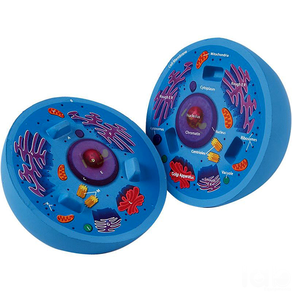
Quick Answer: Animal Cell Model is a one-piece, brightly coloured model of an animal cell on a stand with base, showing the nucleus, endoplasmic reticulum, mitochondria, ribosomes, Golgi apparatus, centrioles, lysosomes and fat vacuoles, numbered with a key card.
Last updated: October 2026
Key Features
Shows the nucleus, endoplasmic reticulum, mitochondria, ribosomes/polysomes and Golgi apparatus
Also shows centrioles, lysosomes and fat vacuoles
Presents the extrusion of a Golgi vesicle and pinocytic signs
Brightly coloured components bring out the segmentation of the cell
One piece, on a stand with base
Numbered, with key card
Technical Specifications
Specification
Value
Construction
One piece
Mounting
Stand with base
Colouring
Brightly coloured components
Labelling
Numbered, key card
About This Product
Bright colouring separates each component of the cell so students can see how finely an animal cell is divided into compartments. Besides the main organelles, the model presents cell dynamics through the extrusion of a Golgi vesicle and pinocytic signs. This one-piece Biology Lab Equipment model stands on a base, with every numbered part listed on the key card.
Frequently Asked Questions
Q1. Which organelles does the Animal Cell Model show? It shows the nucleus, endoplasmic reticulum, mitochondria, ribosomes and polysomes, Golgi apparatus, centrioles, lysosomes and fat vacuoles, with numbered parts on a key card.
Beyond India, Lab Equipments Ambala ships to laboratories and education buyers in Antigua and Barbuda, Bosnia and Herzegovina, Sierra Leone and Belgium.