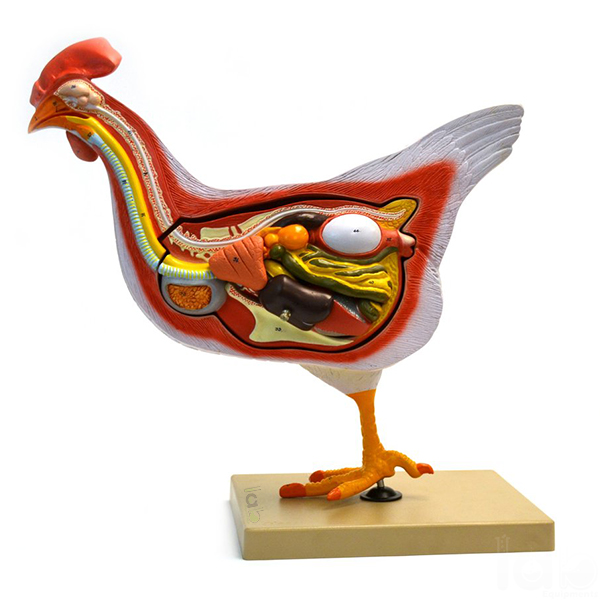
Quick Answer: Hen Anatomy Model is a life-size hen with its left side showing external morphology and its right side longitudinally sectioned to reveal the liver, intestine, stomach, muscles, heart, lungs, reproductive system and an egg.
Last updated: October 2026
Key Features
Life size
Left side shows external morphology
Right side longitudinally sectioned
Inner structures: liver, intestine, stomach, muscles, heart and lungs
Reproductive system shown with an egg
About This Product
By pairing an intact outer side with a sectioned inner side, the model lets students compare the hen's external form with the organs inside the same body. On the right, this life-size Biology Lab Equipment model is sectioned lengthwise to expose the liver, intestine, stomach, muscles, heart and lungs, along with the reproductive system and an egg.
Frequently Asked Questions
Q1. Which organs does the sectioned side show? The longitudinally sectioned right side reveals the liver, intestine, stomach, muscles, heart, lungs, reproductive system and an egg.