
Zoological Models Manufacturer, Suppliers and Exporter in Ambala
Lab Equipments Ambala is leading Zoological Models Manufacturer,and supplier and Exporter in Ambala India, Algeria (Algiers), Angola (Luanda), Argentina (Buenos Aires), Armenia (Yerevan), Australia(Canberra), Austria (Vienna), Bahrain (Manama), Bangladesh (Dhaka), Bhutan (Thimphu), Bolivia (Sucre), Botswana (Gaborone), Brazil (Brasília), Brunei (Bandar Seri Begawan), Montenegro (Podgorica), Morocco (Rabat), Mozambique (Maputo), Myanmar (Naypyidaw), Namibia (Windhoek), Nepal (Kathmandu), New Zealand (Wellington), Nigeria (Abuja), Oman (Muscat), Palestine (Ramallah), Panama (Panama City), Papua New Guinea (Port Moresby), Paraguay (Asunción), Peru (Lima), Philippines (Manila)¸ Portugal (Lisbon), Qatar (Doha), Rwanda (Kigali), Saudi Arabia (Riyadh), Senegal (Dakar), Serbia (Belgrade), Sierra Leone (Freetown), Slovakia (Bratislava), South Africa (Cape Town) (Pretoria) (Bloemfontein), South Sudan (Juba), Spain (Madrid), Sri Lanka (Sri Jayawardenepura Kotte) (Colombo), Sudan (Khartoum), Syria (Damascus), Tanzania (Dodoma), Thailand (Bangkok), Togo (Lomé), Tonga (Nuku'alofa), Trinidad and Tobago (Port of Spain), Tunisia (Tunis), Turkey (Ankara), Turkmenistan (Ashgabat), Uganda (Kampala), United Arab Emirates (Abu Dhabi), United Kingdom (London), United States (Washington, D.C.)
Enlarged and natural-size dissection models of the frog, earthworm, cockroach, rat, fish, hen and pigeon form the core of Zoological Models at Lab Equipments Ambala, used for teaching animal anatomy. Smaller organisms are covered by enlarged models of Hydra, Paramecium, Euglena and the Head of the Pork Tape Worm, and the Frog Development Set shows 24 stages from egg to adult. Cell biology lessons can draw on the meiosis and mitosis model sets and the DNA Model Kit.
Last updated: October 2026
Frequently Asked Questions
Q1. Which models show internal organs after dissection?
The Frog Dissection Model shows external characters dorsally and internal anatomy ventrally, the Bird Dissection - Pigeon Model shows all internal organs at natural size, and the rat, cockroach, fish and earthworm models expose their organ systems.
Q2. How many models are in the cell division sets?
The Animal Cell Division Meiosis Model is a set of 12 models and the Animal Cell Division Mitosis Model a set of 10, with chromosomes painted for easy identification.
Q3. Do the models come with a key card?
Several, including the Bird Dissection - Pigeon Model, Paramecium Model, Locust Anatomy Model and Animal Cell Division Mitosis Model, are numbered with an English key card.
Request a Quote
To add a Frog Dissection Model and the Frog Development Set to your zoology shelf, ask Lab Equipments Ambala.
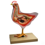
Bird Dissection (Domestic Hen) Model
Product Code : EL-ZM-10592
The right side shows the feathers and all external characters on other side shows
Note:
View Details
Fish Dissection Model
Product Code : EL-ZM-10593
Skeletal structure, brain, digestive tract, swim bladder, reproductive organs and many other anatomical details are clearly exposed.
Note:
View Details
Earthworm Dissection (Pheretima Posthuma) Model
Product Code : EL-ZM-10594
Model showing the external character in a portion and the dissection in the remaining
Note:
View Details
Frog Dissection Model
Product Code : EL-ZM-10595
A beautiful three dimensional model of frog showing all external characters on the dorsal side
Note:
View Details
Bird Dissection - Pigeon Model
Product Code : EL-ZM-10596
Natural Size, Dissection showing all the internal organs of a pigeon.
Note:
View Details
Frog Development Set
Product Code : EL-ZM-10597
A set of 24 models showing the various stages in the development of a frog, from egg to adult.
Note:
View Details
Cockroach Dissection Male Model
Product Code : EL-ZM-10598
An enlarged beautiful model of cockroach dissection (male) showing prothorax
Note:
View Details
Rat Dissection Female Model
Product Code : EL-ZM-10599
An enlarged beautifully coloured model of rat dissection (female) showing head, mouth
Note:
View Details
Ascaris Male and Female Model
Product Code : EL-ZM-10600
Mouth, pharynx, lips, intestine, vagina, female pore, rectum, anus, uterus, oviduct
Note:
View Details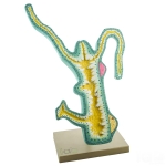
Hydra Model
Product Code : EL-ZM-10601
Colourfully illustrated are the external and internal structure: bud, gastrovascular cavity
Note:
View Details
Animal Cell Division Meiosis Model
Product Code : EL-ZM-10602
A set of 12 models, showing detailed stages in meiotic cell division beautifully coloured
Note:
View Details
Head of the Pork Tape Worm Model
Product Code : EL-ZM-10603
Taenia solium, enlarged many times in one piece showing rostellum, rostral hooks, neck
Note:
View Details
Paramecium Model
Product Code : EL-ZM-10604
Enlarged, dissection showing the structure. Mounted on base.
Note:
View Details
Euglena Model
Product Code : EL-ZM-10605
Enlarged view showing all the important parts like cytostome, stigma, reservoir
Note:
View Details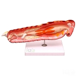
Locust Anatomy Model
Product Code : EL-ZM-10606
Sclistocerca gregaria - The desert Locust. Numbered with English key card.
Note:
View Details
DNA Model Kit
Product Code : EL-ZM-10607
Emphasizing the base pair sequence and function of DNA, the sturdy, colorful bases snap together
Note:
View Details
RNA Model
Product Code : EL-ZM-10608
Model showing RNA molecule DNA Template molecule of chromosome, role of messanger
Note:
View Details
Animal Cell Division Mitosis Model
Product Code : EL-ZM-10609
The chromosomes are painted to allow easy identification, completely numbered with English key card.
Note:
View Details