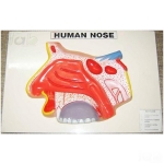
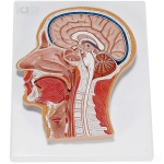
Quick Answer: Anatomical Human Nasal Cavity Model shows the lateral nasal wall with its nasal conchae, the frontal, sphenoid and maxillary paranasal sinuses, and a section of the nasal bones and cartilages of the external nose.
Last updated: October 2026
Key Features
Lateral nasal wall showing the upper, middle and lower nasal conchae projecting medially into the nasal cavity
Paranasal sinuses: frontal, sphenoid and maxillary
External nose sectioned to show the nasal bones and cartilages
About This Product
Seen from the lateral nasal wall, the model shows the upper, middle and lower nasal conchae projecting medially into the nasal cavity. The frontal, sphenoid and maxillary paranasal sinuses are also shown, while a section through the external nose reveals its nasal bones and cartilages. In this way the Head and Facial Model covers the outer nose, the nasal cavity and the paranasal sinuses in one piece.
Frequently Asked Questions
Q1. Which sinuses are shown on the Anatomical Human Nasal Cavity Model? It shows the frontal, sphenoid and maxillary paranasal sinuses.
Beyond India, Lab Equipments Ambala ships to laboratories and education buyers in Honduras, Suriname and Timor-Leste.