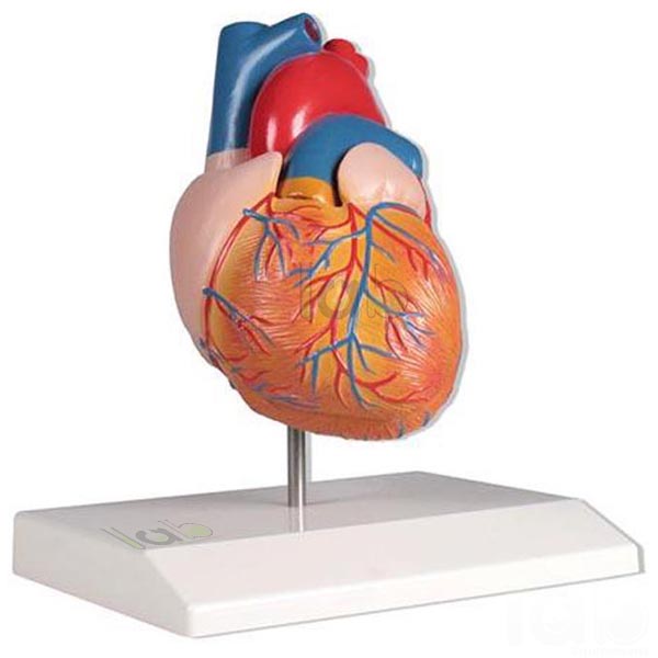
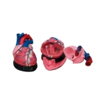
Quick Answer: Human Heart Model is enlarged up to 2–3x and shows the atria with auricles, the coronary sulcus, coronary and pulmonary vessels, and segments of the oesophagus, trachea, aorta and vena cava. Its anterior wall comes off to show the flexible tricuspid, mitral and aortic valves.
Last updated: October 2026
Key Features
Enlarged up to 2–3x the original size
Atria with auricles, coronary sulcus, coronary and pulmonary blood vessels
Segments of the oesophagus, trachea, aorta and vena cava
Removable anterior wall, including the entrance points of both venae cavae
Flexible tricuspid, mitral and aortic valves
Shows a portion of the pericardium; mounted on a base with key card
Technical Specifications
Specification
Value
Enlargement
Up to 2–3x
Removable
Anterior wall
Valves
Flexible tricuspid, mitral and aortic
Chambers
Left auricle and ventricle, right auricle and ventricle
Mounting
Base
Labelling
Numbered, with key card
About This Product
To study the inside of the heart, the anterior wall of this Human System Model is removed, taking with it the entrance points of both venae cavae and opening the way to the flexible tricuspid, mitral and aortic valves. Outside, the auricles, coronary sulcus and a portion of the pericardium are shown.
Frequently Asked Questions
Q1. Are the heart valves visible on the Human Heart Model? Yes. Removing the anterior wall shows the flexible tricuspid, mitral and aortic valves.
Q2. Which vessels are shown? The coronary and pulmonary blood vessels, with segments of the aorta, vena cava, trachea and oesophagus.